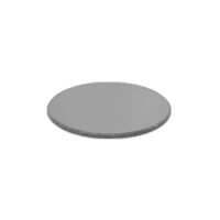
Quick OverviewFilter Color: Matte Finish White. Filter Size:
Dia. 32mm. For BM0503, BM0504, BM0505, BM1401, BM1403, BM1404, BM1307, BM1303, BM1304, PL1302, BM0301, NIKON-E100 Series Microscope. FI02013101 32mm Filter (Matte Finish White)Color FilterFilter ColorMatte Finish WhiteFilter Size Dia. 32mmMaterialGlassNet Weight0.002kg (0.004lbs)Applied FieldFor BM0503, BM0504, BM0505, BM1401, BM1403, BM1404, BM1307, BM1303, BM1304, PL1302, BM0301, NIKON-E100 Series Microscope
.tb_part
{
width: 100%;
border: solid 0px #ccc;
font-size:12px;
}
.tb_part tr
{
height: 30px;
line-height: 30px;
}
.tb_part td
{
white-space: nowrap;
}
.tb_part_1 td
{
white-space: wrap;
}
.normal{
border-left:solid 2px black;
border-right:solid 2px black;
}
.tb_part tr.normal td
{
border:solid 1px #ccc;
padding-right:5px;
}
.tb_part tr.normal:nth-child(2){
border-top:solid 2px black;
}
.tb_part tr.normal:first-child{
border:none;
}
.tb_part tr.normal:first-child td{
border:none;
font-weight:bold;
font-size:16px;
}
.end-border{
border-bottom:solid 2px black;
}
.tb_part tr.memo,.tb_part tr.memo td{
border:none;
}
Technical InfoInstructionsIlluminatorClose ΛThe conditions of different illumination of the microscope are a very important parameter. Choosing the correct illumination method can improve the resolution and contrast of the image, which is very important for observing the imaging of different objects.
The wavelength of the light source is the most important factor affecting the resolution of the microscope. The wavelength of the light source must be smaller than the distance between the two points to be observed in order to be distinguished by the human eye. The resolution of the microscope is inversely proportional to the wavelength of the light source. Within the range of the visible light, the violet wavelength is the shortest, providing also the highest resolution. The wavelength of visible light is between 380~780nm, the maximum multiple of optical magnification is 1000-2000X, and the limit resolution of optical microscope is about 200nms. In order to be able to observe a much smaller object and increase the resolution of the microscope, it is necessary to use light having a much shorter wavelength as the light source. The most commonly used technical parameters for describing illumination are luminescence intensity and color temperature. Luminescence intensity, with lumen as unit, is the physical unit of luminous flux. The more lumens, the stronger the illumination. Color temperature, with K (Kelvin) as unit, is a unit of measure indicating the color component of the light. The color temperature of red is the lowest, then orange, yellow, white, and blue, all gradually increased, with the color temperature of blue being the highest. The light color of the incandescent lamp is warm white, its color temperature is 2700K, the color temperature of the halogen lamp is about 3000K, and the color temperature of the daylight fluorescent lamp is 6000K.
A complex and complete lighting system can include a light source, a lampshade or lamp compartment, a condenser lens, a diaphragm, a variety of wavelength filters, a heat sink cooling system, a power supply, and a dimming device etc. Select and use different parts as needed. Of which, selection and use of the illuminating light source is the most important part of the microscope illumination system, as and other components are designed around the illuminating wavelength curve and characteristics of the illuminating light source.
Some of the microscope light sources are pre-installed on the body or frame of the microscope, and some are independent. There are many types and shapes of light sources. Depending on the requirements of the microscope and the object to be observed, one type or multiple types of illumination at the same time can be selected. In addition, the whole beam and band adjustment of the light source, the position and illumination angle of the light source, and the intensity and brightness of the light all have a great influence on the imaging. For microscope imaging, a good lighting system may be a system that allows for more freedom of adjustment. In actual work, such as industry, too many adjustment mechanisms may affect the efficiency of use, therefore choose the appropriated configured lighting conditions is very important. Color FilterClose ΛColor filter is a type of filter that allows light of only a certain wavelength and color range to pass, while light of other wavelengths is intercepted. Color filter is made of colored glass, and it has various bandwidths and color for selection. Both artificial light source (lamp light) and natural light (daylight) are all full-color light, including seven colors, namely, red, orange, yellow, green, blue, indigo and purple. As the microscope illumination, different types of light sources have different color temperatures and brightness. In order to adjust the color of the light source, it is necessary to install a filtering device at the light exit port of the light source, so that the spectrum of a certain wavelength band is transmitted or blocked. Color filter generally can only be added to the illumination path to change the color of the illumination source and improve the contrast of the image, but generally it is not installed in the imaging path system, which affect the image quality.
There are many types of color filters. In addition to the color requirements, color filters of different colors also contribute to the imaging quality. Color filters using the same color will brighten the color of the image.
Of the traditional daylight filter, there are relatively more red and yellow light in the lamp light, the resolution is not high, and the observation is not comfortable. The use of daylight filter can absorb the color between yellow to red spectrum emitted by the light source, thus the color temperature becomes much closer to daylight, making microscope observation more comfortable, and it is one of the most used microscope color filters. Daylight blue filter can get close to the daylight spectrum, obtain more short-wave illumination, and improve the resolution of the objective lens. For example, using blue color filter (λ=0.44 microns) can improve the resolution by 25% than green color filter (λ=0.55 microns). Therefore, blue color filter can improve the resolution, and improve the image effect observed under the microscope. However, the human eye is sensitive to green light with a wavelength of about 0.55 microns. When using blue color filters for photomicrography, it is often not easy to focus on the projection screen. Yellow and green filters: both yellow and green filters can increase the contrast (i.e. contrast ratio) of details of the specimen. As far as the achromatic objective lens is concerned, the aberrations in the yellow and green bands are better corrected. Therefore, when yellow and green color filters are used, only yellow and green light passes, and the aberration will be reduced, thereby improving the imaging quality. For semi-apochromatic and apochromat objectives, the focus of visible light is concentrated. In principle, any color filter can be used, but if yellow and green filters are used, the color will make the human eye feel comfortable and soft. Red filter. Red has the longest wavelength and the lowest resolution in visible light. However, red light image can filter and eliminate the variegated background in the image. Therefore, so it has a very good effect for some applications that do not require color features for identification, and the edges and contours of the image are also the clearest, which is more accurate for measurement. Medium gray filters, also known as neural density filters, or ND for short, can uniformly reduce visible light. It is suitable for photomicrography and connection to computer monitors for observation. ND can be used for exposure control and good light absorption, and reduce the light intensity while not changing the color temperature of the microscope light source. PackagingClose ΛAfter unpacking, carefully inspect the various random accessories and parts in the package to avoid omissions. In order to save space and ensure safety of components, some components will be placed outside the inner packaging box, so be careful of their inspection. For special packaging, it is generally after opening the box, all packaging boxes, protective foam, plastic bags should be kept for a period of time. If there is a problem during the return period, you can return or exchange the original. After the return period (usually 10-30 days, according to the manufacturer’s Instruction of Terms of Service), these packaging boxes may be disposed of if there is no problem.
About Boli Optics:
Boli Optics Microscope Supplier sells professional microscopes, microscope accessories, and magnifying lamps. We offer parts and accessories compatible with Leica, Olympus, Nikon, and Zeiss, and more. We supply research laboratories, medical centers, universities, industrial manufactures, factories, and more. Our engineers and technicians provide technical support and design & manufacture custom microscope products for your applications. Our products are manufactured under ISO 9001 quality control standards. We also provide OEM service. Since 1994, our talented team has been working in the optics industry and serving our customers whole-heartedly. Based in Southern California, we offer fast, same day shipping from our local warehouses.
Visit Product PageEmail:
sales@bolioptics.com
Location:
8762 Lanyard Court,
Rancho Cucamonga
California
91730


