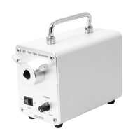
Instruction ManualML212112 Series LED Fiber Optic Illuminator.pdfQuick OverviewLED Light. Light Adjustable. Fiber Cable Adapter Size: 5/8 in.
End Adapter. Pointer Panel Meter/Scale. Output Power: 50W. Input Voltage: AC 100-240V 50/60Hz. Output Voltage: DC 24V. Power Cord Connector Type: USA 2 Pins. ML21211311 50W LED Fiber Optic IlluminatorFiber Optic Light SourceLight Source TypeLED LightPower Supply AdjustableLight AdjustableLight Source Illuminance50000Lux or MorePower Box Light PortSingle HoleFiber Cable Adapter Size5/8 in.
End AdapterPower Box Panel Meter DisplayPointer Panel Meter/ScalePower Box Cooling SystemFan Cooling systemPower Box Dimensions176x100x130mmBulb Color Temperature6400KSingle Bulb Luminous Flux≥400LMOutput Power50WInput VoltageAC 100-240V 50/60HzOutput VoltageDC 24VPower Cord Connector TypeUSA 2 PinsPower Cable Length1.8mSurface TreatmentSpray PaintMaterialMetalColorWhiteNet Weight3.00kg (6.61lbs)
.tb_part
{
width: 100%;
border: solid 0px #ccc;
font-size:12px;
}
.tb_part tr
{
height: 30px;
line-height: 30px;
}
.tb_part td
{
white-space: nowrap;
}
.tb_part_1 td
{
white-space: wrap;
}
.normal{
border-left:solid 2px black;
border-right:solid 2px black;
}
.tb_part tr.normal td
{
border:solid 1px #ccc;
padding-right:5px;
}
.tb_part tr.normal:nth-child(2){
border-top:solid 2px black;
}
.tb_part tr.normal:first-child{
border:none;
}
.tb_part tr.normal:first-child td{
border:none;
font-weight:bold;
font-size:16px;
}
.end-border{
border-bottom:solid 2px black;
}
.tb_part tr.memo,.tb_part tr.memo td{
border:none;
}
Technical InfoInstructionsIlluminatorClose ΛThe conditions of different illumination of the microscope are a very important parameter. Choosing the correct illumination method can improve the resolution and contrast of the image, which is very important for observing the imaging of different objects.
The wavelength of the light source is the most important factor affecting the resolution of the microscope. The wavelength of the light source must be smaller than the distance between the two points to be observed in order to be distinguished by the human eye. The resolution of the microscope is inversely proportional to the wavelength of the light source. Within the range of the visible light, the violet wavelength is the shortest, providing also the highest resolution. The wavelength of visible light is between 380~780nm, the maximum multiple of optical magnification is 1000-2000X, and the limit resolution of optical microscope is about 200nms. In order to be able to observe a much smaller object and increase the resolution of the microscope, it is necessary to use light having a much shorter wavelength as the light source. The most commonly used technical parameters for describing illumination are luminescence intensity and color temperature. Luminescence intensity, with lumen as unit, is the physical unit of luminous flux. The more lumens, the stronger the illumination. Color temperature, with K (Kelvin) as unit, is a unit of measure indicating the color component of the light. The color temperature of red is the lowest, then orange, yellow, white, and blue, all gradually increased, with the color temperature of blue being the highest. The light color of the incandescent lamp is warm white, its color temperature is 2700K, the color temperature of the halogen lamp is about 3000K, and the color temperature of the daylight fluorescent lamp is 6000K.
A complex and complete lighting system can include a light source, a lampshade or lamp compartment, a condenser lens, a diaphragm, a variety of wavelength filters, a heat sink cooling system, a power supply, and a dimming device etc. Select and use different parts as needed. Of which, selection and use of the illuminating light source is the most important part of the microscope illumination system, as and other components are designed around the illuminating wavelength curve and characteristics of the illuminating light source.
Some of the microscope light sources are pre-installed on the body or frame of the microscope, and some are independent. There are many types and shapes of light sources. Depending on the requirements of the microscope and the object to be observed, one type or multiple types of illumination at the same time can be selected. In addition, the whole beam and band adjustment of the light source, the position and illumination angle of the light source, and the intensity and brightness of the light all have a great influence on the imaging. For microscope imaging, a good lighting system may be a system that allows for more freedom of adjustment. In actual work, such as industry, too many adjustment mechanisms may affect the efficiency of use, therefore choose the appropriated configured lighting conditions is very important. Fiber Optic Light SourceClose ΛFiber optic light source refers to an illuminating light source that does not contain or contains less spectrum of infrared heat radiation in a illuminating or light guiding body, for example, the popular LED light source, which is a typical illuminator fiber optic light source. In microscopic illumination, the optical fiber cold light source (commonly referred to as “cool light”) means that, after the illumination beam is transmitted through the optical fiber of the light guide body, the heat radiation is not brought to the light exit port, thereby achieving "cold light" effect.
The portion of the illuminating light source of the optical fiber has been conventionally illuminated with a halogen light source. In recent years, high-power LED lighting has been widely used. Although the bulb of halogen light source can generate a lot of heat radiation, because of its high brightness when emitting light, it belongs to full-band light, with good color reproduction and comfortable observation by human eye, and therefore is still irreplaceable in some applications. Luminous light sources usually require a high-power light source to achieve strong light, therefore heat dissipation is very important. Whether it is a halogen light source or an LED light source, fan cooling is usually adopted.
Fiber optic lighting application has many advantages: 1. The thermal conductivity of the optical fiber is poor. When the light source (light bulb) emits light, the thermal radiation, after being separated by the optical fiber, is not transmitted to the object to be observed. So, while maintaining the wavelength and brightness of the light, it becomes "cold light". When using strong light, cold light may not damage the observed objects, especially in medical and biological applications. 2. Single light source can be transmitted through the optical fiber, and at the same time there are multiple light-emitting points with the same light-emitting characteristics. The light-emitting port can be arranged at different positions and angles, or made into different shapes, such as double-branch lighting, ring lighting, multi-point lighting etc. 3. The light source host and the light exit port illumination point are transmitted through the optical fiber, and therefore the host can be placed in a safe or suitable position without affecting the illumination position of the light exit port, so that there will be more flexibility in design and use. 4. The light exiting port illumination point is transmitted through the optical fiber, and it can filter freely the wavelength of the light at the light source position in the front end of the light entrance, increase the polarization effect, and adjust the brightness and darkness. For example, improve the contrast and contrast ratio of the details of the object to be observed through various color filters, filter out the ultraviolet and infrared light, and reduce damage to certain items.. 5. In the light source host and optical fiber used in fiber optic lighting, the service life of the optical fiber can be decades, and the design separating the light source from the optical fiber makes the light source easy to repair and replace. Light AdjustableClose ΛThe brightness of the light source adjustable is very important in the imaging of the microscope. Since the difference of the numerical aperture of the objective lens of high magnification and low magnification is very big, more incident light is needed to achieve a much better resolution when using a high magnification objective lens. Therefore, when observing through a high magnification objective lens, the brightness required is high; when observing through a low magnification objective lens, the brightness required is low. When observing different objects, or feature points of the same object at different positions, the brightness needs are also different; including the difference of background light or reflection within the field of view of observation, it has a great influence on the effect of observing the object, and therefore one needs to adjust the brightness of the light source according to each object to be observed. In the light source capable of providing continuous spectrum, such as a halogen lamp, the brightness adjustment of the light not only adjusts the brightness and intensity of the light, but also changes the spectrum emitted by the light source. When the light source is dark, there are many components of red light, and when the brightness is high, there are more blue spectrum. If the required light is strong and the spectrum needs to be changed, the light can be kept at a brighter intensity, which is solved by adjusting the spectrum by adding a color filter.
Take note of the dimming button on the light source, after the On/Off switch is turned on, normally clockwise is to brighten, and counterclockwise is to darken.
If it is adjusted to the lowest brightness, the light source should normally be lit. If the naked eye still can't see the object being illuminated brightly, you need to adjust the brightness knob to a much bigger position. Generally, there is scale marking on the dimming knob, which is an imaginary number representing the percentage of brightness, or an electronic digital display, giving the brightness of the light source under the same conditions a marking. PackagingClose ΛAfter unpacking, carefully inspect the various random accessories and parts in the package to avoid omissions. In order to save space and ensure safety of components, some components will be placed outside the inner packaging box, so be careful of their inspection. For special packaging, it is generally after opening the box, all packaging boxes, protective foam, plastic bags should be kept for a period of time. If there is a problem during the return period, you can return or exchange the original. After the return period (usually 10-30 days, according to the manufacturer’s Instruction of Terms of Service), these packaging boxes may be disposed of if there is no problem.
About Boli Optics:
Boli Optics Microscope Supplier sells professional microscopes, microscope accessories, and magnifying lamps. We offer parts and accessories compatible with Leica, Olympus, Nikon, and Zeiss, and more. We supply research laboratories, medical centers, universities, industrial manufactures, factories, and more. Our engineers and technicians provide technical support and design & manufacture custom microscope products for your applications. Our products are manufactured under ISO 9001 quality control standards. We also provide OEM service. Since 1994, our talented team has been working in the optics industry and serving our customers whole-heartedly. Based in Southern California, we offer fast, same day shipping from our local warehouses.
Visit Product PageEmail:
sales@bolioptics.com
Location:
8762 Lanyard Court,
Rancho Cucamonga
California
91730


