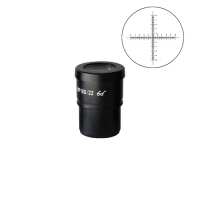
Quick OverviewReticle Eyepiece. Eyepiece Optical Magnification: 10X. Eyepiece Size for Eye Tube:
Dia. 30mm. Eyepiece Field of View:
Dia. 22mm. Crosshair Scale. Reticle Dimensions:
Dia. 24x1.5mm. 10mm/100 Div . SZ05013232 10X Reticle Eyepiece ( Dia. 30/FN22)EyepieceEyepiece TypeReticle EyepieceEyepiece Optical Magnification10XPlan EyepiecePlan EyepieceEyepiece Size for Eye Tube Dia. 30mmEyepiece Field of View Dia. 22mmEyepoint TypeHigh Eyepoint EyepieceEyepiece Size for Reticle Dia. 24mmReticle TypeCrosshair ScaleReticle Dimensions Dia. 24x1.5mmScale Range10mm/100 Div Division Value0.1mmSurface TreatmentElectroplating BlackMaterialMetalColorBlackNet Weight0.07kg (0.15lbs)
.tb_part
{
width: 100%;
border: solid 0px #ccc;
font-size:12px;
}
.tb_part tr
{
height: 30px;
line-height: 30px;
}
.tb_part td
{
white-space: nowrap;
}
.tb_part_1 td
{
white-space: wrap;
}
.normal{
border-left:solid 2px black;
border-right:solid 2px black;
}
.tb_part tr.normal td
{
border:solid 1px #ccc;
padding-right:5px;
}
.tb_part tr.normal:nth-child(2){
border-top:solid 2px black;
}
.tb_part tr.normal:first-child{
border:none;
}
.tb_part tr.normal:first-child td{
border:none;
font-weight:bold;
font-size:16px;
}
.end-border{
border-bottom:solid 2px black;
}
.tb_part tr.memo,.tb_part tr.memo td{
border:none;
}
Technical InfoInstructionsEyepieceClose ΛEyepiece is also called ocular.
Eyepiece is a visual optical device that is used to observe an image formed by an objective lens after magnification. Eyepiece can magnify the real image obtained by the magnification of the objective lens to form a clear and erect virtual image at the least distance of distinct vision (LDDV). For aberration, eyepiece is usually composed of several lenses that correct the residual aberrations produced during imaging by the objective lens. Usually, eyepiece has a relatively larger field of view and angular magnification.
The eyepiece is provided with a diaphragm in the middle of the upper and lower lenses, or at the lower end of the lower lens, wherein micrometer, reticle, pointer and other accessories are mounted.
On the eyepiece there is engraved with the following marks: eyepiece type, magnification, field of view number, and the like. For example, eyepiece is engraved with PL10X/22; PL represents plane eyepiece, or flat-field eyepiece, 10X is the magnification, 22 represents the field of view, and the glasses mark represents high eyepoint eyepiece, and C represents compensating eyepiece. Generally, the outer diameter of the eyepiece of a compound microscope is 23.2 mm, and stereo microscope can have a much larger field of view diameter because of its low magnification, so the outer diameter is mostly 30 mm (or 30.5 mm).
The resolution capability of the microscope is determined by the numerical aperture of the objective lens, while the eyepiece plays only the magnification role. Therefore, for structures and details that cannot be distinguished by the objective lens, the eyepiece still cannot distinguish even if it is magnified again. Reticle EyepieceClose ΛReticle eyepiece. The eyepiece focal length (10mm below the eyepiece mounting surface eyepiece shoulder) of the reticle eyepiece is equipped with a reticle for measuring and positioning the object to be observed. For one microscope, a reticle can be installed only on one eyepiece, and it requires that the two eyepieces should be completely identical. If two are installed, it is generally very difficult to make the two reticles of both the left and the right eyepiece completely overlap, which may cause eye discomfort. The reticle eyepieces are generally used on 10X and 20X eyepieces. The mounting dimension of the reticle refers to the size of the inner diameter of the lower end tube of the eyepiece. It requires that the eyepiece that can be equipped with a reticle needs a preset thread and a pressing ring. The reticle is facing up (in the direction of the eyepiece lens), placed flat on the reticle mounting surface of the eyepiece, and screwed in with a pressure ring, and press tight. Ordinary users can also install the reticle on their own as needed. The reticle is generally made of glass material, and the etched printed surface is the front side. It is mounted on the end close to the eye, which is the position of the eyepiece image plane; when avoiding the use of different reticle, the focal length is different due to the different thickness of the glass, which makes the scribe line of the reticle fall on the unclear image plane position.
The reticle is placed under the eyepiece. When measuring the object, the reticle and the object to be measured are also magnified by the eyepiece, so the actual length of the reading has no relation at all with the magnification of the eyepiece. When the reticle reads the length value in the eyepiece of the microscope, because the length of the image to be measured passes through the objective lens and reaches the image plane position of the reticle, the length read is actually the length magnified by the objective lens. The real numerical value should be the length of the reading, divided by the numerical value of magnification of the objective lens. If it is the zoom microscope body with also magnification, it should also be divided by the magnification (objective lens X zoom). In this measurement method, the error mainly lies in that the magnification of the objective lens is not calibrated, and the magnification error of ordinary microscope objective lens can reach +-5%. Therefore, for accurate measurement, it should be used after calibration with the objective micrometer. For the calibration method, please refer to the introduction of “Reticle”. Eyepiece Optical MagnificationClose ΛEyepiece optical magnification is the visual magnification of the virtual image after initial imaging through the eyepiece. When the human eye observes through the eyepiece, the ratio of the tangent of the angle of view of the image and the tangent of the angle of view of the human eye when viewing or observing the object directly at the reference viewing distance is usually calculated according to 250 mm/focal length of eyepiece. The standard configuration of a general microscope is a 10X eyepiece. Usually, the magnification of the eyepiece of compound microscope is 5X, 8X, 10X, 12.5X, 16X, 20X. As stereo microscope has a low total magnification, its eyepiece magnification generally does not use 5X, but can achieve 25X, 30X and other much bigger magnification. Eyepiece Field of ViewClose ΛThe eyepiece field of view is the diameter of the field diaphragm of the eyepiece, or the diameter of the image plane of the field diaphragm imaged by the field diaphragm. The diameter of a large field of view can increase the viewing range, and see more detail in the field of view. However, if the field of view is too large, the spherical aberration and distortion around the eyepiece will increase, and the stray light around the field of view will affect the imaging effect. Eyepoint TypeClose ΛEye point refers to the axial distance between the upper end of the metal frame of the eyepiece and the exit of pupil. The exit of pupil distance of high eyepoint eyepiece is farther than that of the eye lens of the ordinary eyepiece. When this distance is greater than or equal to 18mm, it is a high eyepoint eyepiece. When observing, one does not need to be too close to the eyepiece lens, making it comfort to observe, and it can also be viewed with glasses. Generally, there is a glasses logo on the eyepiece, indicating that it is a high eyepoint eyepiece. PackagingClose ΛAfter unpacking, carefully inspect the various random accessories and parts in the package to avoid omissions. In order to save space and ensure safety of components, some components will be placed outside the inner packaging box, so be careful of their inspection. For special packaging, it is generally after opening the box, all packaging boxes, protective foam, plastic bags should be kept for a period of time. If there is a problem during the return period, you can return or exchange the original. After the return period (usually 10-30 days, according to the manufacturer’s Instruction of Terms of Service), these packaging boxes may be disposed of if there is no problem.
About Boli Optics:
Boli Optics Microscope Supplier sells professional microscopes, microscope accessories, and magnifying lamps. We offer parts and accessories compatible with Leica, Olympus, Nikon, and Zeiss, and more. We supply research laboratories, medical centers, universities, industrial manufactures, factories, and more. Our engineers and technicians provide technical support and design & manufacture custom microscope products for your applications. Our products are manufactured under ISO 9001 quality control standards. We also provide OEM service. Since 1994, our talented team has been working in the optics industry and serving our customers whole-heartedly. Based in Southern California, we offer fast, same day shipping from our local warehouses.
Visit Product PageEmail:
sales@bolioptics.com
Location:
8762 Lanyard Court,
Rancho Cucamonga
California
91730